D2 is the major source of T3, especially in the brain
In humans, approximately 70% of circulating T3 comes from D2, 15% from D1 and the rest from the thyroid. In hypothyroidism D2 activity increases and D1 activity reduces. Hence D2 is crucial and especially so in hypothyroidism. In addition to circulating T3, D2 also supplies intracellular T3. D2 is the major source of T3 in the brain. Thus, the major factor that regulates thyroid status in the brain is the regulation of local T3 levels by D2 and D3.
Gereben B, McAninch EA, Ribeiro MO, Bianco AC. Scope and limitations of iodothyronine deiodinases in hypothyroidism. Nat Rev Endocrinol. 2015 Nov;11(11):642-652. doi: 10.1038/nrendo.2015.155. Epub 2015 Sep 29. PMID: 26416219; PMCID: PMC5003781.
A study in rats demonstrated that more than half the T3 bound to nuclear receptors in the brain comes from conversion of T4 to T3, in the cerebral cortex almost 80% of T3 is derived from T4 locally.
D2 has the dominant role in determining thyroid hormone status in local tissues
T3 saturation levels in the cell nucleus determine thyroid status: whether the tissue will be euthyroid, hypothyroid or hyperthyroid. Intracellular T3 levels are determined by a combination of circulating T3 plus T3 derived from D2 regulated by cell-specific factors (such as the response to cold exposure in BAT). In tissues that express D2 it has a major role in regulating T3. T3 from D2 activity is produced close to the cell nucleus and ‘hangs around’, as opposed to circulating T3 from the thyroid or D1 activity. In a way there are ‘two T3s’: T3 and what we might call ‘D2T3’. Although exactly the same, ‘D2T3’ has a crucial role in cellular thyroid status and hence the clinical status of the patient.
Abdalla SM, Bianco AC. Defending plasma T3 is a biological priority. Clin Endocrinol (Oxf). 2014 Nov;81(5):633-41. doi: 10.1111/cen.12538. Epub 2014 Aug 7. PMID: 25040645; PMCID: PMC4699302.
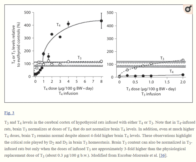
T3 and T4 in the brain
The major cellular transport proteins, OATP1C1 and MCT8, favour T4 over T3. Hence T4 crosses the blood brain barrier (BBB) in preference to T3. Within the brain D2 and D3 regulate T3 levels.
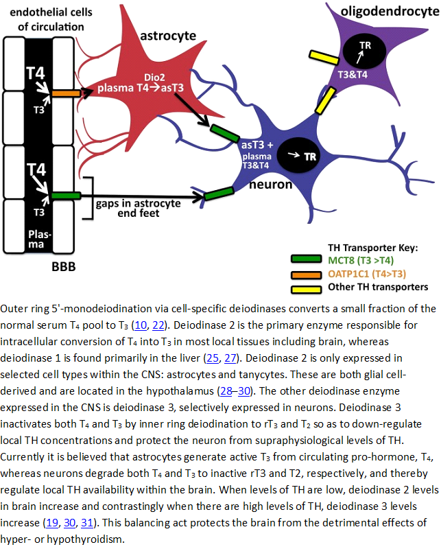
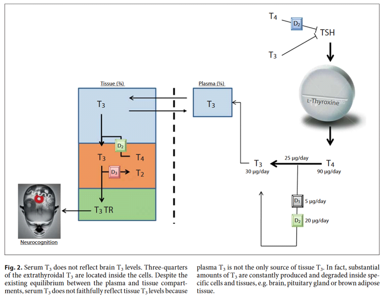
It’s important to note that serum T3 does not accurately reflect brain (and other tissue) T3 levels. Most T3 is inside the cells, not in the serum and is subject to local regulation by D2 and D3.
Bianco AC, Casula S. Thyroid hormone replacement therapy: three ‘simple’ questions, complex answers. Eur Thyroid J. 2012 Jul;1(2):88-98. doi: 10.1159/000339447. Epub 2012 Jun 27. PMID: 24783002; PMCID: PMC3821470.
The following figure from the same paper shows how critical D2 and D3 activity is. In this experiment on rats you can see from the left-hand graph that brain T3 stays within normal limits even when T4 (solid circles) is well below or well above normal levels. On the other hand, brain T3 is normalized only if 3x a replacement T3 dose is given. This is consistent with my personal experience; I need to take around 40 mcg L-T3 before normal cognitive function is restored. Although I can’t measure my brain T3 levels I have found that the right amount of T3 restores normal sleep patterns with adequate deep sleep and cognitive function during the day. This raises a dilemma because adequate T3 for normal brain function may be too much for other organs.
Bianco AC, Casula S. Thyroid hormone replacement therapy: three ‘simple’ questions, complex answers. Eur Thyroid J. 2012 Jul;1(2):88-98. doi: 10.1159/000339447. Epub 2012 Jun 27. PMID: 24783002; PMCID: PMC3821470.
D2 activity in the brain is crucial, it’s clear that anything that impairs D2 activity will have major consequences. We now look at the evidence that TSH Regulates Deiodinase and in particular D2.